Treating Mortons Neuroma

Overview

plantar neuromaA Morton?s Neuroma is actually incorrectly termed, with the name suggesting it is a tumour or growth. Rather than a true neuroma it is actually what is called a perineural fibrosis, which means that over time the sheath surrounding the nerve becomes irritated, inflamed, and forms a thickened scar tissue.

Causes

A Morton’s neuroma commonly occurs due to repetitive weight bearing activity (such as walking or running) particularly when combined with tight fitting shoes or excessive pronation of the feet (i.e. “flat-feet”). The condition is also more common in patients with an unstable forefoot allowing excessive movement between the metatarsal bones. A Morton’s neuroma can also occur due to certain foot deformities, trauma to the foot, or the presence of a ganglion or inflamed bursa in the region which may place compressive forces on the nerve.

Symptoms

The symptoms of Morton?s Neuroma tend to come and go over time. They are typically exacerbated by physical activity or by wearing certain shoes. Morton?s Neuroma symptoms include sharp pain in the ball of the foot, pain radiating to the tips of the toes, burning pain in the second, third, or fourth toes, numbness in the toes, sensation of a lump between the toes.

Diagnosis

The doctor will ask about your symptoms and medical history. A physical exam will be done. Initial diagnosis of Morton’s neuroma is based on your description of the type and location of pain and discomfort in the foot. The diagnosis will be confirmed by a physical exam of the foot, including checking for mechanical abnormalities in the foot, squeezing the side of the foot, which will usually cause pain when Morton’s neuroma is present. Examination of your shoes to check for excess wear in parts of the shoe, check to see whether the shoes are too tight. Imaging tests evaluate the foot and surrounding structures. This may be done with X-ray, MRI scan, Ultrasound. Injections of local anesthetic can also be used for diagnosis.

Non Surgical Treatment

Once a diagnosis is obtained, it is essential to begin treatment immediately. Your podiatric physician will advise you on the most effective means. If caught early enough, good foot care, shoes that fit properly, and/or orthoses may eliminate the need for any further intervention. Other conservative measures might include oral non-steroidal anti-inflammatory medication (NSAIDS), physical therapy, ultrasound or other non-invasive measures. If that doesn?t work, your podiatric physician might use injectable steroids, and/or a local anesthetic around the neuroma to reduce inflammation and pain. Many patients report relief after these measures are taken.interdigital neuroma

Surgical Treatment

If these non-surgical measures do not work, surgery is sometimes needed. Surgery normally involves a small incision (cut) being made on either the top, or the sole, of the foot between the affected toes. Usually, the surgeon will then either create more space around the affected nerve (known as nerve decompression) or will cut out (resect) the affected nerve. If the nerve is resected, there will be some permanent numbness of the skin between the affected toes. This does not usually cause any problems. You will usually have to wear a special shoe for a short time after surgery until the wound has healed and normal footwear can be used again. Surgery is usually successful. However, as with any surgical operation, there is a risk of complications. For example, after this operation a small number of people can develop a wound infection. Another complication may be long-term thickening of the skin (callus formation) on the sole of the foot (known as plantar keratosis). This may require treatment by a specialist in care of the feet (chiropody).

Prevention

How can Morton?s neuroma be prevented? Do not wear tight shoes or high-heeled shoes for prolonged periods. Do wear shoes with a wide toe box so that your toes are not squeezed or cramped. Do wear athletic footwear with enough padding to cushion the balls of the feet when exercising or participating in sports.

A Way To Identify Heel Spur

Heel Spur

Overview

A heel spur is a deposit of calcium on the heel bone. This calcification takes the form of a bony protrusion, which can cause considerable pain when standing and walking. This foot problem is closely related to plantar fasciitis, a condition in which the band of fibrous tissue on the bottom of the foot becomes over-stressed. It pulls away from the heel and causes the calcium deposits to form. For this reason, treating a heel spur involves treating the plantar fascia as well.

Causes

Heel spurs develop as an abnormal growth in the heel bone due to calcium deposits that form when the plantar fascia pulls away from the heel. This stretching of the plantar fascia is usually the result of over-pronation (flat feet), but people with unusually high arches (pes cavus) can also develop heel spurs. Women have a significantly higher incidence of heel spurs due to the types of footwear often worn on a regular basis.

Inferior Calcaneal Spur

Symptoms

Heel spur and plantar fasciitis pain usually begins in the bottom of the heel, and frequently radiates into the arch. At times, however, the pain may be felt only in the arch. The pain is most intense when first standing, after any period of rest. Most people with this problem experience their greatest pain in the morning, with the first few steps after sleeping. After several minutes of walking, the pain usually becomes less intense and may disappear completely, only to return later with prolonged walking or standing. If a nerve is irritated due to the swollen plantar fascia, this pain may radiate into the ankle. In the early stages of Heel Spurs and Plantar Fasciitis, the pain will usually subside quickly with getting off of the foot and resting. As the disease progresses, it may take longer periods of time for the pain to subside.

Diagnosis

Most patients who are suffering with heel spurs can see them with an X-ray scan. They are normally hooked and extend into the heel. Some people who have heel spur may not even have noticeable symptoms, although could still be able to see a spur in an X-ray scan.

Non Surgical Treatment

In extreme cases, a doctor may recommend surgery for the removal of heel spurs. Fortunately, this is the exception rather than the rule. Most cases can be resolved with a combination of icing, rest, foot stretches and supporting the foot with an orthodic shoe insert specifically designed for this condition. We recommend that you continue on to our article on Heel Spur Treatment to discover the best, speediest and most affordable methods of resolving this ailment without invasive medical procedures.

Surgical Treatment

More than 90 percent of people get better with nonsurgical treatments. If conservative treatment fails to treat symptoms of heel spurs after a period of 9 to 12 months, surgery may be necessary to relieve pain and restore mobility. Surgical techniques include release of the plantar fascia, removal of a spur. Pre-surgical tests or exams are required to identify optimal candidates, and it’s important to observe post-surgical recommendations concerning rest, ice, compression, elevation of the foot, and when to place weight on the operated foot. In some cases, it may be necessary for patients to use bandages, splints, casts, surgical shoes, crutches, or canes after surgery. Possible complications of heel surgery include nerve pain, recurrent heel pain, permanent numbness of the area, infection, and scarring. In addition, with plantar fascia release, there is risk of instability, foot cramps, stress fracture, and tendinitis.

Prevention

Use orthotic inserts. You can purchase orthotics over the counter, or you can have orthotics specially fitted by your podiatrist. Try 1 of these options. Heel cups. These inserts will help to align the bones in your foot and to cushion your heel. Check your skin for blisters when you first start using heel cups. Also, your feet may sweat more with a heel cup, so change your socks and shoes often. Insoles. While you can pick up generic insoles at a drugstore, you may have more luck if you go to a store that sells athletic shoes. Push on the arch to make sure that it doesn’t collapse. If your insoles help but could use a little work, you can take them to a podiatrist to get them customized. Custom orthotics. A podiatrist can make a cast of your foot and provide you with custom-made orthotics. These may be more expensive, but they are made of materials specifically designed for your needs, and they can last up to 5 years if your podiatrist refurbishes them every 1 or 2 years. To find a podiatrist near you, look at the Web page for the American Academy of Podiatric Sports Medicine. Dynamic Insoles. Lack of elasticity in plantar fascia in the foot is for most people the real problem. If there is poor elasticity in the lengthwise tendons in the foot (plantar fascia) in relation to a person’s general condition, only a small additional strain is required for the pull on the tendons to cause damage to the tissues connecting the tendons to the heel bone. This will generate an inflamed condition called Plantar Fasciitis.

What Is The Perfect Strategy For Inferior Calcaneal Spur

Calcaneal Spur

Overview

It shouldn’t hurt to get to your feet in the morning or walk throughout your day, but if your steps result in stabbing or aching pain in one or both heels, you may be suffering from heel spurs. Also known as calcaneal spurs or osteophytes, heel spurs are pointed, hooked or shelf-shaped calcium build-ups on the heel bone (calcaneus). While the spurs, themselves, do not sense pain, their tendency to prod the soft, fatty tissues of the heel can result in severe discomfort with every step you take. This article will teach you what you need to know about heel spurs so that you can understand your symptoms and find fast relief from your pain.

Causes

The pain caused by heel spurs can be a sharp, stabbing pain when using the foot after a long period of rest. Sometimes it then reduces to a dull throb that can worsen when engaging in activities like jogging or jumping. People sometimes describe the pain of heel spurs and plantar fasciitis as a pin sticking into the bottom of the foot when they first stand up in the morning, this pain later turns into a bearable ache. The cause of the pain is generally not the heel spur itself, but the soft-tissue buildup associated with it. People often complain that the sharp pain returns after they stand up following sitting for a prolonged period of time.

Heel Spur

Symptoms

An individual with the lower legs turning inward, a condition called genu valgus or “knock knees,” can have a tendency toward excessive pronation. This can lead to a fallen arch and problems with the plantar fascia and heel spurs. Women tend to suffer from this condition more than men. Heel spurs can also result from an abnormally high arch. Other factors leading to heel spurs include a sudden increase in daily activities, an increase in weight, or a thinner cushion on the bottom of the heel due to old age. A significant increase in training intensity or duration may cause inflammation of the plantar fascia. High-heeled shoes, improperly fitted shoes, and shoes that are too flexible in the middle of the arch or bend before the toe joints will cause problems with the plantar fascia and possibly lead to heel spurs.

Diagnosis

Because the diagnosis of heel spurs can be confused with tarsal tunnel syndrome (as described earlier), most surgeons advocate performing a tarsal tunnel release (or at least a partial tarsal tunnel release) along with the plantar fascia release. This surgery is about 80percent successful in relieving pain in the small group of patients who do not improve with conservative treatments.

Non Surgical Treatment

Heel spurs can be solved with simple solutions that do not involve surgery. Avoiding extended periods of activity such as running, sports and walking. Applying ice directly to the heel for 5 minutes at a time. This helps soothe and reduce inflammation. Lose weight to reduce stress on your heels. A series of simple exercises. Inexpensive orthotic shoe inserts. The best way to treat heel spurs is by treating the underlying cause of the problem. This involves correcting the dynamics of your foot motion with orthotic insoles.

Surgical Treatment

In some cases, heel spurs are removed by surgery after an X-ray. While the surgery is typically effective, it?s a timely and expensive procedure. Even after surgery, heel spurs can re-form if the patient continues the lifestyle that led to the problem. These reasons are why most people who develop painful heel spurs begin looking for natural remedies for joint and bone pain. Surgery isn?t required to cure a heel spur. In fact, more than 90 percent of people get better with nonsurgical treatments. If nonsurgical methods fail to treat symptoms of heel spurs after 12 months, surgery may be necessary to alleviate pain and restore mobility.

Prevention

In 2002, researchers attempted to compare the effects of various running techniques on pronation and resulting injuries like stress fractures and heel spurs. They suggested that it is possible to teach runners to stride in such a way as to minimize impact forces. One way is to lower running speed. Another is to take longer rest periods following a run.

Bursitis Top Of Foot Treatment Plan

Overview

Retrocalcaneal bursitis is sometimes difficult to differentiate from Achilles tendinitis, at least symptomatically. Both are most uncomfortable during the push-off phase of gait, are most severely painful in the morning and with walking after sitting for a period of time, and generally worsen with activity. Most practitioners make the distinction between the two simply on the basis of location of pain and tenderness. Generally, Achilles tendinitis is felt an inch or two higher than this form of bursitis.

Causes

There are several factors which can lead to a person developing retrocalcaneal bursitis. In athletes, especially runners, overtraining, sudden excessive increase in running mileage may lead to retrocalcaneal bursitis. Tight or ill-fitting shoes can be another causative factor as they can produce excessive pressure at the back of the heel due to restrictive heel counter. A person with an excessively prominent posterosuperior aspect of the heel bone (Haglund deformity) may also have a higher predisposition to retrocalcaneal bursitis. In such individuals, pain would be reproduced when the ankle goes into dorsiflexion.

Symptoms

Your heel may feel more sensitive to the cold and ache in cold and damp weather due to impaired circulation. These symptoms are often the result of failure to treat the injury properly from the outset and overicing.

Diagnosis

Gram stain. A lab test called a Gram stain is used to determine if certain troublesome bacteria are present. Not all bacteria can be identified with a Gram stain, however, so even if the test comes back negative, septic bursitis cannot be completely ruled out. White blood cell count. An elevated number of white blood cells in the bursa’s synovial fluid indicates an infection. Glucose levels test. Glucose levels that are significantly lower than normal may indicate infection.

Non Surgical Treatment

Orthotics may assist heel bursitis by providing stability to the heel, reduce any foot functioning abnormalities and provide extra support for the feet. The orthotic achieves this by maintaining correct foot posture, therefore facilitating normal functioning of the Achilles tendon. Icing the back of the heel post activity for temporary relief. Non-steroidal anti-inflammatory medication such as ibuprofen can reduce inflammation of the bursa. Stretching of the calf muscle may reduce the pulling on the heel by the Achilles tendon. Shoes that have an elevated heel may reduce pulling on the heel from the Achilles tendon. Resting the painful heel may reduce inflammation and pain. Surgical removal of the painful bursa is a last resort treatment when all other treatments have failed.

Surgical Treatment

Bursectomy is a surgical procedure used to remove an inflamed or infected bursa, which is a fluid-filled sac that reduces friction between tissues of the body. Because retrocalcaneal bursitis can cause chronic inflammation, pain and discomfort, bursectomy may be used as a treatment for the condition when it is persistent and cannot be relived with other treatments. During this procedure, a surgeon makes small incisions so that a camera may be inserted into the joint. This camera is called an arthroscope. Another small incision is made so that surgical instruments can be inserted to remove the inflamed bursa.

Hammer Toe

Hammer ToeOverview

Many disorders can affect the joints in the toes, causing pain and preventing the foot from functioning as it should. A hammertoes occurs when the joint at the end of the toe cannot straighten. Excessive rubbing of the hammer toe against the top of the shoe can lead to pain and the development of a corn. The tip of the toe is often turned down against the shoe causing pressure and discomfort.

Causes

Footwear can contribute significantly to the development of hammertoes. Shoes that are too small force your toes into a curled position. Over time, your toe tendons adjust to this positioning, causing your toe or toes to hold a hammered shape. Athletes may be especially susceptible, because of the increased forces on the toes from shoes that are too small or tight. Heel elevation in footwear is also problematic, as it causes your toes to be pushed into the shoe?s toe box. Heel elevation additionally contributes to muscle imbalance. A common example of this is when your Achilles tendon-the tendon at the back of your leg that attaches your calf muscles to your heel bone-is too tight, causing the tendons on the top of your foot that attach to your toes to work too hard and hold your toes in an unnatural, elevated position.

Hammer ToeSymptoms

People who have painful hammertoes visit their podiatrist because their affected toe is either rubbing on the end their shoe (signaling a contracted flexor tendon), rubbing on the top of their shoe (signaling a contracted extensor tendon), or rubbing on another toe and causing a painful buildup of thick skin, known as a corn.

Diagnosis

Most health care professionals can diagnose hammertoe simply by examining your toes and feet. X-rays of the feet are not needed to diagnose hammertoe, but they may be useful to look for signs of some types of arthritis (such as rheumatoid arthritis) or other disorders that can cause hammertoe hammertoe.

Non Surgical Treatment

Putting padding between your toes and strapping them in place can help to stop pain caused by the toes rubbing. Custom-made insoles for your shoes will help to take the pressure off any painful areas. Special shoes that are wider and deeper than normal can stop your toes rubbing. However if your pain persists your consultant may recommend an surgery.

Surgical Treatment

In advanced cases in which the toe has become stiff and permanently bent, the toe can be straightened with surgery. One type of surgery involves removing a small section of the toe bone to allow the toe to lie flat. Surgery for hammertoe usually is classified as a cosmetic procedure. Cosmetic foot surgeries sometimes cause complications such as pain or numbness, so it?s better to treat the problem with a shoe that fits properly.

HammertoePrevention

The best treatment is good prevention! Hammertoe can be prevented by wearing shoes with ample toe room, avoiding high heels, and wearing adjustable shoes to assure a looser fit. When buying shoes, shop at the end of the day when your feet are swollen from daily activity, try both shoes on to confirm they fit properly, and if necessary, visit a shoe repair store to see if they can stretch your shoes for a better fit.

What Actually Causes Feet To Over Pronate

Overview

n a normal functioning ankle and foot there should be 15- to 20-degrees of ankle dorsiflexion present (leg moving forward on top of the foot; or, same as the foot bending backward toward the leg) as well as 60- to 65-degrees of first toe extension (bending backward). Foot pronation (turning downward or inward) and supination (turning upward or outward) are normal and needed movements for proper function during the gait cycle. All feet must do this and do it well in order to minimize the forces that the body has to deal with during walking or running, and also to create the lever that is needed for propulsion.Over-Pronation

Causes

A common cause of pronation is heredity – we can inherit this biomechanical defect. The second most common cause is due to the way our feet were positioned in the uterus while we were developing; this is called a congenital defect. In either instance, the following occurs in our feet during our development.

Symptoms

Due to the laxity of the soft tissue structures of the foot, and the fact that the joints are not held together properly, the bones of the feet shift. When this occurs, the muscles that attach to these bones must also shift, or twist, in order to attach to these bones. The strongest and most important muscles that attach to our foot bones come from our lower leg. So, as these muscles course down the leg and across the ankle, they must twist to maintain their proper attachments in the foot. This twisting of these muscles will cause shin splints, Achilles Tendonitis, generalized tendonitis, fatigue, muscle aches and pains, cramps, ankle sprains, and loss of muscular efficiency (reducing walking and running speed and endurance). The problems we see in the feet, which are due to over-pronation include bunions, heel spurs, plantar fasciitis, fallen and painful arches, hammertoes, metatarsalgia (ball of foot pain), and calluses.

Diagnosis

Your healthcare provider will ask about your symptoms, medical history, and activities and examine your feet. Your provider may watch you walk or run. Check the motion of your feet when they strike the ground. Look at your athletic shoes to see if they show an abnormal pattern of wear.Over Pronation

Non Surgical Treatment

Over-Pronation can be treated conservatively (non-surgical treatments) with over-the-counter orthotics. These orthotics should be designed with appropriate arch support and medial rearfoot posting to prevent the over-pronation. Footwear should also be examined to ensure there is a proper fit. Footwear with a firm heel counter is often recommended for extra support and stability. Improper fitting footwear can lead to additional problems of the foot. If the problem persists, call your doctor to schedule an appointment.

Surgical Treatment

Calcaneal “Slide” (Sliding Calcaneal Osteotomy) A wedge is cut into the heel bone (calcaneus) and a fixation device (screws, plate) is used to hold the bone in its new position. This is an aggressive option with a prolonged period of non-weightbearing, long recovery times and many potential complications. However, it can and has provided for successful patient outcomes.

Calcaneal Apophysitis In Youngsters

Overview

Sever’s disease is a condition characterized by pain in one or both heels with walking. The pain is caused by shortening of the heel-cord. It usually affects children between the ages of 10 and 13 years old. During this phase of life, growth of the bone is taking place at a faster rate than the tendons. Sever’s disease is also called calcaneal apophysitis.

Causes

Sever?s disease is directly related to overuse of the bone and tendons in the heel. This can come from playing sports or anything that involves a lot of heel movement. It can be associated with starting a new sport, or the start of a new season. Children who are going through adolescence are also at risk of getting it because the heel bone grows quicker than the leg. Too much weight bearing on the heel can also cause it, as can excessive traction since the bones and tendons are still developing. It occurs more commonly in children who over-pronate, and involves both heels in more than half of patients.

Symptoms

The most obvious sign of Sever’s disease is pain or tenderness in one or both heels, usually at the back. The pain also might extend to the sides and bottom of the heel, ending near the arch of the foot. A child also may have these related problems, swelling and redness in the heel, difficulty walking, discomfort or stiffness in the feet upon awaking, discomfort when the heel is squeezed on both sides, an unusual walk, such as walking with a limp or on tiptoes to avoid putting pressure on the heel. Symptoms are usually worse during or after activity and get better with rest.

Diagnosis

Sever’s disease is based on the symptoms reported. To confirm the diagnosis, the clinician will examine the heels and ask about the child’s activity level and participation in sports. They may also squeeze the back part of the heel from both sides at the same time to see if doing so causes pain and also ask the child to stand on tiptoes to see if that position causes pain. There may be tightness in the calf muscle, which contributes to tension on the heel. Symptoms are usually worse during or after activity and get better with rest. X-rays generally are not that helpful in diagnosing Sever’s disease, but they may be ordered to rule out other problems, such as fractures. Sever’s disease cannot be seen on an X-ray.

Non Surgical Treatment

The initial treatment is rest, relieve the pain and treat the underlying cause. The foot and ankle should be rested so that the apophysis it is not being continually ?injured?. A small heel (sorbothane heel insert) raise can used to raise the heel and take the stretch off the Achilles tendon. It may be sufficient to stop your child playing sport but some children require a short period of bed or couch rest. Some children find resting very difficult and require the use of braces or plasters or boots to slow them down. Using crutches is advised. Severs is usually caused by tight muscles. A stretching program should be followed usually supervised by a physiotherapist. The stretching program may need to be undertaken up to 5 times a day. If flat feet are a problems orthotics (insoles) should be used. The pain should be controlled by rest (limiting activity) and ice (icing the painful area 3-4 times a day – making sure the skin is not burnt), Simple pain killers can be used such as paracetamol as well as anti-inflammatory tablets and cream. Severs disease usually goes away with time. When your child stops growing, the pain and swelling should go away because the growing (weak) area fuses and becomes solid bone which is very strong.

Surgical Treatment

The surgeon may select one or more of the following options to treat calcaneal apophysitis. Reduce activity. The child needs to reduce or stop any activity that causes pain. Support the heel. Temporary shoe inserts or custom orthotic devices may provide support for the heel. Medications. Nonsteroidal anti-inflammatory drugs (NSAIDs), such as ibuprofen, help reduce the pain and inflammation. Physical therapy. Stretching or physical therapy modalities are sometimes used to promote healing of the inflamed issue. Immobilization. In some severe cases of pediatric heel pain, a cast may be used to promote healing while keeping the foot and ankle totally immobile. Often heel pain in children returns after it has been treated because the heel bone is still growing. Recurrence of heel pain may be a sign of calcaneal apophysitis, or it may indicate a different problem. If your child has a repeat bout of heel pain, be sure to make an appointment with your foot and ankle surgeon.

Understanding Heel Ache

Overview

Painful Heel

Plantar Fasciitis, also known as Heel Pain or a heel spur, is a musculoskeletal condition causing pain under the heel or into the inner arch of the foot. The condition is commonly mistaken for an impact trauma or heel bruise but in fact it is caused by mechanical overstretching of the fibrous tissue in the arch. Heel pain can develop suddenly or evolve gradually over time. It can affect people of all ages, but is more common beyond the 4th decade of life, those in standing occupations, overweight individuals and those involved in regular strenuous exercise.

Causes

Plantar fasciitis can come from a number of underlying causes. Finding the precise reason for the heel pain is sometimes difficult. As you can imagine, when the foot is on the ground a tremendous amount of force (the full weight of the body) is concentrated on the plantar fascia. This force stretches the plantar fascia as the arch of the foot tries to flatten from the weight of your body. This is just how the string on a bow is stretched by the force of the bow trying to straighten. This leads to stress on the plantar fascia where it attaches to the heel bone. Small tears of the fascia can result. These tears are normally repaired by the body. As this process of injury and repair repeats itself over and over again, a bone spur (a pointed outgrowth of the bone) sometimes forms as the body’s response to try to firmly attach the fascia to the heelbone. This appears on an X-ray of the foot as a heel spur. Bone spurs occur along with plantar fasciitis but they are not the cause of the problem. As we age, the very important fat pad that makes up the fleshy portion of the heel becomes thinner and degenerates (starts to break down). This can lead to inadequate padding on the heel. With less of a protective pad on the heel, there is a reduced amount of shock absorption. These are additional factors that might lead to plantar fasciitis. Some physicians feel that the small nerves that travel under the plantar fascia on their way to the forefoot become irritated and may contribute to the pain. But some studies have been able to show that pain from compression of the nerve is different from plantar fasciitis pain. In many cases, the actual source of the painful heel may not be defined clearly. Other factors that may contribute to the development of plantar fasciitis include obesity, trauma, weak plantar flexor muscles, excessive foot pronation (flat foot) or other alignment problems in the foot and or ankle, and poor footwear.

Symptoms

The symptoms of plantar fasciitis include pain along the inside edge of the heel near the arch of the foot. The pain is worse when weight is placed on the foot especially after a long period of rest or inactivity. This is usually most pronounced in the morning when the foot is first placed on the floor. This symptom called first-step pain is typical of plantar fasciitis. Prolonged standing can also increase the painful symptoms. It may feel better after activity but most patients report increased pain by the end of the day. Pressing on this part of the heel causes tenderness. Pulling the toes back toward the face can be very painful.

Diagnosis

After you have described your foot symptoms, your doctor will want to know more details about your pain, your medical history and lifestyle, including. Whether your pain is worse at specific times of the day or after specific activities. Any recent injury to the area. Your medical and orthopedic history, especially any history of diabetes, arthritis or injury to your foot or leg. Your age and occupation. Your recreational activities, including sports and exercise programs. The type of shoes you usually wear, how well they fit, and how frequently you buy a new pair. Your doctor will examine you, including. An evaluation of your gait. While you are barefoot, your doctor will ask you to stand still and to walk in order to evaluate how your foot moves as you walk. An examination of your feet. Your doctor may compare your feet for any differences between them. Then your doctor may examine your painful foot for signs of tenderness, swelling, discoloration, muscle weakness and decreased range of motion. A neurological examination. The nerves and muscles may be evaluated by checking strength, sensation and reflexes. In addition to examining you, your health care professional may want to examine your shoes. Signs of excessive wear in certain parts of a shoe can provide valuable clues to problems in the way you walk and poor bone alignment. Depending on the results of your physical examination, you may need foot X-rays or other diagnostic tests.

Non Surgical Treatment

Physical medicine modalities are well known for their benefits and they have been consistently applied in early treatment of plantar fasciitis. Typically, the direct application of ice, ice baths or contrast soaking aid in the local reduction of inflammation and temporarily augment pain management. Electric stimulation may only provide indirect reduction of interstitial inflammation of the plantar fascia. Ultrasound therapy, hot pack systems and deep tissue massage help eliminate inflammation and aid in restoring plantar fascia tensegrity. Generally, these modalities are considered to be valuable adjuncts to a well-organised treatment plan. Various programs of stretching, range of motion and therapeutic exercises can help re-establish foot function and improve tolerance to load. When it is done appropriately, stretching can serve as an important adjunct to the resumption of the plantar fascia?s ability to tolerate eccentric loading forces that typically occur during stance and gait. Night splinting has proven to be an effective tool in managing persistent plantar fasciitis. Antiinflammatory modalities, such as ice and ice baths, are often the first line of treatment. Oral NSAIDs have been a mainstay of treatment. While they effectively relieve symptoms, be aware that they frequently fail to promote sustained relief. When inflammation is severe or fails to respond to initial efforts, one may consider corticosteroid injection(s). However, keep in mind that corticosteroid injections impose the risk of aponeurosis rupture secondary to focal collagen tissue necrosis and can result in focal heel fat pad atrophy.

Surgical Treatment

Surgery is a last resort in the treatment of heel pain. Physicians have developed many procedures in the last 100 years to try to cure heel pain. Most procedures that are commonly used today focus on several areas, remove the bone spur (if one is present), release the plantar fascia (plantar fasciotomy), release pressure on the small nerves in the area. Usually the procedure is done through a small incision on the inside edge of the foot, although some surgeons now perform this type of surgery using an endoscope. An endoscope is a tiny TV camera that can be inserted into a joint or under the skin to allow the surgeon to see the structures involved in the surgery. By using the endoscope, a surgeon can complete the surgery with a smaller incision and presumably less damage to normal tissues. It is unclear whether an endoscopic procedure for this condition is better than the traditional small incision. Surgery usually involves identifying the area where the plantar fascia attaches to the heel and releasing the fascia partially from the bone. If a small spur is present this is removed. The small nerves that travel under the plantar fascia are identified and released from anything that seems to be causing pressure on the nerves. This surgery can usually be done on an outpatient basis. This means you can leave the hospital the same day.

Prevention

Heel Discomfort

Make sure you wear appropriate supportive shoes. Don’t over-train in sports. Make sure you warm up, cool down and undertake an exercise regime that helps maintain flexibility. Manage your weight, obesity is a factor in causing plantar fasciitis. Avoid walking and running on hard surfaces if you are prone to pain. You should follow the recognized management protocol “RICED” rest, ice, compression, elevation and diagnosis. Rest, keep off the injured ankle as much as possible. Ice, applied for 20 minutes at a time every hour as long as swelling persists. Compression, support the ankle and foot with a firmly (not tightly) wrapped elastic bandage. Elevation, keep foot above heart level to minimize bruising and swelling. Diagnosis. Consult a medical professional (such as a Podiatrist or doctor) especially if you are worried about the injury, or if the pain or swelling gets worse. If the pain or swelling has not gone down significantly within 48 hours, also seek treatment. An accurate diagnosis is essential for proper rehabilitation of moderate to severe injuries.

Posted in Heel Pain

Tags:

Permalink

The Leading Causes And Treatment Plans Of Achilles Tendon Pain

Overview

Achilles Tendinitis
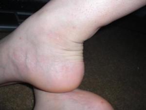
The Achilles tendon, which connects the calf muscles to the heel bone, can withstand significant pressure from physical activities. Achilles tendinitis is estimated to account for approximately 11 percent of all running injuries, as the Achilles tendon provides the momentum to push off to walk or run. Achilles tendinitis, also called Achilles tendinopathy, results from overuse, injury or disease of the Achilles tendon, which causes the area to become inflamed. There are two types of Achilles tendinitis: Non-insertional Achilles Tendinitis – Fibers that are located in the middle portion of the tendon began to develop small tears that cause swelling and thickening. This type of tendinitis is usually found in younger people who are very active. Insertional Achilles Tendinitis – Develops where the tendon attaches to the heel bone in the lower part of the heel. Extra bone growth also called bone spurs form because of this tendinitis and can affect patients at any time, even if they are not active.

Causes

Tight or fatigued calf muscles, which transfer the burden of running to the Achilles. This can be due to poor stretching, rapidly increasing distance, or over-training excessive hill running or speed work, both of which stress the Achilles more than other types of running. Inflexible running shoes, which, in some cases, may force the Achilles to twist. Runners who overpronate (feet rotate too far inward on impact) are most susceptible to Achilles tendinitis.

Symptoms

Recurring localized pain, sometimes severe, along the tendon during or a few hours after running. Morning tenderness about an inch and a half above the point where the Achilles tendon is attached to the heel bone. Sluggishness in your leg. Mild or severe swelling. Stiffness that generally diminishes as the tendon warms up with use.

Diagnosis

Physicians usually pinch your Achilles tendon with their fingers to test for swelling and pain. If the tendon itself is inflamed, your physician may be able to feel warmth and swelling around the tissue, or, in chronic cases, lumps of scar tissue. You will probably be asked to walk around the exam room so your physician can examine your stride. To check for complete rupture of the tendon, your physician may perform the Thompson test. Your physician squeezes your calf; if your Achilles is not torn, the foot will point downward. If your Achilles is torn, the foot will remain in the same position. Should your physician require a closer look, these imaging tests may be performed. X-rays taken from different angles may be used to rule out other problems, such as ankle fractures. MRI (magnetic resonance imaging) uses magnetic waves to create pictures of your ankle that let physicians more clearly look at the tendons surrounding your ankle joint.

Nonsurgical Treatment

The best treatment for Achilles tendonitis is preventative, stretching and warming up properly before starting an activity. Proper rest, accompanied by stretching and icing to reduce swelling, can help to heal an overworked Achilles tendon. Placing an adequate heel lift in both shoes will allow the heel to have contact with the ground without placing stress on the Achilles tendon. Wear a tie shoe that is stiff soled and has a wide base, then add an over-the-counter or custom foot orthosis inside the shoe to prevent the twisting motion of the Achilles tendon due to over pronation. In the event that the tendon is unable to heal due to your life style or activity, you may have to be put in a walking cast for a short period to give it a chance to heal. You need to have the doctor, physical therapist, or come in to our facility to check for a leg length difference due to the walking cast being higher. This is to prevent any discomfort to the hips. After the tendon has healed and before the foot is taken out of the walking cast, range of motion at the ankle must be tested and if the foot is not allowed to properly bend upwards 15 degrees then the tightness in the calf will cause the foot to over pronate and reinjure. A stretching program will be needed to loosen up the calf muscle before much weight bearing is done without the cast. The stretching program can be found at the menu for feet hurt. If necessary a heel lift can be put in both shoes to help take the stress off the tendon. Should the tightness of the calves be the primary cause for the Achilles tendon damage and stretching has not loosen the Achilles tendon sufficiently, then discussion with your doctor for a calf release may have to be considered.

Achilles Tendonitis

Surgical Treatment

Most people will improve with simple measures or physiotherapy. A small number continue to have major problems which interfere with their lifestyle. In this situation an operation may be considered. If an operation is being considered, the surgeon will interview you and examine you again and may want you to have further treatment before making a decision about an operation. Before undergoing Achilles tendonitis surgery, London based patients, and those who can travel, will be advised to undergo a scan, which will reveal whether there is a problem in the tendon which can be corrected by surgery. Patients will also have the opportunity to ask any questions and raise any concerns that they may have, so that they can proceed with the treatment with peace of mind.

Prevention

While it may not be possible to prevent Achilles tendinitis, you can take measures to reduce your risk. Increase your activity level gradually. If you’re just beginning an exercise regimen, start slowly and gradually increase the duration and intensity of the training. Take it easy. Avoid activities that place excessive stress on your tendons, such as hill running. If you participate in a strenuous activity, warm up first by exercising at a slower pace. If you notice pain during a particular exercise, stop and rest. Choose your shoes carefully. The shoes you wear while exercising should provide adequate cushioning for your heel and should have a firm arch support to help reduce the tension in the Achilles tendon. Replace your worn-out shoes. If your shoes are in good condition but don’t support your feet, try arch supports in both shoes. Stretch daily. Take the time to stretch your calf muscles and Achilles tendon in the morning, before exercise and after exercise to maintain flexibility. This is especially important to avoid a recurrence of Achilles tendinitis. Strengthen your calf muscles. Strong calf muscles enable the calf and Achilles tendon to better handle the stresses they encounter with activity and exercise. Cross-train. Alternate high-impact activities, such as running and jumping, with low-impact activities, such as cycling and swimming.

What Leads To Pain At The Heel

Foot Pain

Overview

Plantar fasciitis is a painful inflammatory process of the plantar fascia, a thick fibrous band of connective tissue originating on the bottom surface of the calcaneus (heel bone) and extending along the sole of the foot towards the five toes. Pain in the arch or heel often indicates inflammation of the long band of tissue under the foot (the plantar fascia). It can cause sharp pain and discomfort in either the mid arch region or at the inside heel, and less commonly the outside heel. It frequently causes pain upon rising from rest (especially first thing in the morning) and can progress to agony by the end of the day. Although plantar fasciitis is the most common cause of this pain, it must be skilfully differentially diagnosed from other conditions via a thorough history taking and physical examination.


Causes

Identified risk factors for plantar fasciitis include excessive running, standing on hard surfaces for prolonged periods of time, high arches of the feet, the presence of a leg length inequality, and flat feet. The tendency of flat feet to excessively roll inward during walking or running makes them more susceptible to plantar fasciitis. Obesity is seen in 70% of individuals who present with plantar fasciitis and is an independent risk factor. Studies have suggested a strong association exists between an increased body mass index and the development of plantar fasciitis. Achilles tendon tightness and inappropriate footwear have also been identified as significant risk factors.


Symptoms

Plantar fasciitis is characterized by the following signs and symptoms. Acute plantar fasciitis, pain is usually worse in the morning but may improve when activity continues; if the plantar fasciitis is severe, activity will exacerbate the pain, pain will worsen during the day and may radiate to calf or forefoot, pain may be described anywhere from “minor pulling” sensation, to “burning”, or to “knife-like”, the plantar fascia may be taut or thickened, passive stretching of the plantar fascia or the patient standing on their toes may exacerbate symptoms, acute tenderness deep in the heel-pad along the insertion of the plantar aponeurosis at the medial calcaneal tuberosity and along the length of the plantar fascia, may have localized swelling. Chronic plantar fasciitis, plantar fasciitis is classified as “chronic” if it has not resolved after six months, pain occurs more distally along the aponeurosis and spreads into the Achilles tendon.


Diagnosis

Plantar fasciosis is confirmed if firm thumb pressure applied to the calcaneus when the foot is dorsiflexed elicits pain. Fascial pain along the plantar medial border of the fascia may also be present. If findings are equivocal, demonstration of a heel spur on x-ray may support the diagnosis; however, absence does not rule out the diagnosis, and visible spurs are not generally the cause of symptoms. Also, infrequently, calcaneal spurs appear ill defined on x-ray, exhibiting fluffy new bone formation, suggesting spondyloarthropathy (eg, ankylosing spondylitis, reactive arthritis. If an acute fascial tear is suspected, MRI is done.


Non Surgical Treatment

Treatment of heel pain caused by plantar fasciitis begins with simple steps. There are a number of options for treatment of plantar fasciitis, and almost always some focused effort with nonsurgical treatments can provide excellent relief. In rare circumstances, simple steps are not adequate at providing relief, and more invasive treatments may be recommended. Typically, patients progress from simple steps, and gradually more invasive treatments, and only rarely is surgery required.

Painful Heel


Surgical Treatment

If you consider surgery, your original diagnosis should be confirmed by the surgeon first. In addition, supporting diagnostic evidence (such as nerve-conduction studies) should be gathered to rule out nerve entrapment, particularly of the first branch of the lateral plantar nerve and the medial plantar nerve. Blood tests should consist of an erythrocyte sedimentation rate (ESR), rheumatoid factor, human leukocyte antigen B27 (HLA-B27), and uric acid. It’s important to understand that surgical treatment of bone spurs rarely improves plantar fasciitis pain. And surgery for plantar fasciitis can cause secondary complications-a troubling condition known as lateral column syndrome.


Prevention

Warm up properly. This means not only stretching prior to a given athletic event, but a gradual rather than sudden increase in volume and intensity over the course of the training season. A frequent cause of plantar fasciitis is a sudden increase of activity without suitable preparation. Avoid activities that cause pain. Running on steep terrain, excessively hard or soft ground, etc can cause unnatural biomechanical strain to the foot, resulting in pain. This is generally a sign of stress leading to injury and should be curtailed or discontinued. Shoes, arch support. Athletic demands placed on the feet, particularly during running events, are extreme. Injury results when supportive structures in the foot have been taxed beyond their recovery capacity. Full support of the feet in well-fitting footwear reduces the likelihood of injury. Rest and rehabilitation. Probably the most important curative therapy for cases of plantar fasciitis is thorough rest. The injured athlete must be prepared to wait out the necessary healing phase, avoiding temptation to return prematurely to athletic activity. Strengthening exercises. Below are two simple strength exercises to help condition the muscles, tendons and joints around the foot and ankle. Plantar Rolling, Place a small tin can or tennis ball under the arch of the affected foot. Slowly move the foot back and forth allowing the tin can or tennis ball to roll around under the arch. This activity will help to stretch, strengthen and massage the affected area. Toe Walking, Stand upright in bare feet and rise up onto the toes and front of the foot. Balance in this position and walk forward in slow, small steps. Maintain an upright, balanced posture, staying as high as possible with each step. Complete three sets of the exercise, with a short break in between sets, for a total of 20 meters.